Comprehensive Aortic Program
Program
The evolution of aortic surgery at Northwestern University Feinberg School of Medicine extends across five decades, from before the founding of the Division of Vascular Surgery in the mid-1970s, through the endovascular revolution of the 1980s, 1990s, and 2000s, to the rise of the multidisciplinary and increasingly minimally invasive approach to treating aortic disease today. The Comprehensive Aortic Program (CAP) of the Northwestern Division of Vascular Surgery (NDVS) builds on our longstanding commitment to excellence in aortic surgery and the treatment of a broad range of vascular diseases. With alignment of the broad resources within Northwestern Medicine, we offer comprehensive treatment of all facets of aortic disease in a patient-centered fashion.
Aortic disease has long served as a proving ground for surgical innovation. Northwestern's contributions span open surgical technique, the noninvasive vascular laboratory, endovascular therapy, industry-partnered device development, advanced imaging, and the training of generations of vascular and cardiac surgeons who have gone on to lead programs across the country. A theme that emerges clearly from the historical record, and even more distinctly from the experiences recounted below, is that this progress was never the work of a single specialty or a single “great pioneer.” From the outset, it was a collaborative altruistic effort between many individuals across multiple specialties and backgrounds so close that, as one participant remarked, it is difficult to say where one discipline's contribution ended and the other's began. Collaboration has been a critical element of our success in the clinical treatment of aortic disease. This includes close collaboration with cardiac surgery, cardiology, cardiovascular genetics, radiology and multiple other services. These collaborations enable the delivery of the full spectrum of medical and surgical treatment for aortic disease as well as continued program expansion in a rich educational and research setting.
The robust environment of innovation and collaboration are critical to the delivery of exceptional quality, innovation and personalized patient care. This is the hallmark of the NDVS - CAP.
Aortic History at Northwestern: Past, Present, and Future
The Past: Founding a Division, Founding a Field
The Bergan-Yao Era
Northwestern's modern vascular surgery program traces its roots to the early 1970s, when John J. Bergan, MD, then chief of the Division of Transplantation, recruited James S. T. Yao, MD, PhD, to Northwestern. Dr. Yao established a blood flow laboratory at Northwestern Memorial Hospital in 1972, one of the first noninvasive vascular laboratories in the country. Two years later, Drs. Bergan and Yao created one of the earliest vascular surgery fellowship programs in the United States. In 1976, they formally established the Division of Vascular Surgery within the Department of Surgery, with Dr. Bergan appointed its inaugural chief.
Following Dr. Bergan’s tenure, Dr. Yao served as division chief from 1988 to 1997 and later chaired the Department of Surgery until 2000. He formalized protocols for noninvasive vascular testing and co-founded the Society for Noninvasive Vascular Technology, but his most enduring legacy may be the generations of trainees he and Dr. Bergan mentored, many of whom went on to lead vascular divisions, departments, and medical schools of their own.
Aortic Disease Becomes a Research Focus
William H. Pearce, MD, who succeeded Dr. Yao as division chief in 1998, built an active basic-science program studying the pathogenesis of abdominal aortic aneurysms. This advanced Northwestern within the broader national effort to understand aneurysm biology rather than treat aortic aneurysms as a purely mechanical problem. A landmark 1990 paper by Pearce and colleagues in the American Journal of Pathology used antibody staining to identify T-cell and B-cell infiltration in aneurysm tissue, among the earlier observations establishing that chronic inflammation, not merely mechanical wall degeneration, is central to aneurysm pathology; companion Northwestern papers on collagen composition (1989) and elastin degradation (1992) rounded out a sustained, multi-year program on aneurysm wall biology. Pearce later established the NIH-funded Vascular Surgery Scientist Training Program in 2009, extending the division's research mission into a formal pathway for training vascular surgeon-scientists that remains ongoing.
Early Open Surgery: Precedents and Firsts
Dr. William H. Pearce offered his own interview, detailing the division's open surgical period that predates what the literature readily captures. A bibliography of landmark papers he compiled for this chapter documents that Thomas E. Starzl, MD, in the stretch of his career preceding his far better-known work in transplantation, published on a large aortoiliac endarterectomy at Northwestern in 1959, more than a decade before the division's formal founding. The same bibliography shows that John Bergan himself published on aortic occlusive disease with O. H. Trippel as early as 1963. Trippel's name appears again in a paper on reinforced Ivalon (polyvinyl alcohol) sponge as an aortic graft material, one of the earliest synthetic graft substitutes tried, years before Dacron and Gore-Tex became standard. Pearce also credited Dr. William Flynn, a partner of Bergan and Yao's, with placing what he believes was the first aortic Gore-Tex graft anywhere, a case never formally published.
A Parallel Origin Story: Interventional Radiology and the Angioplasty Revolution
No account of aortic surgery at Northwestern is complete without the parallel story of interventional radiology, recounted by Robert Vogelzang, MD. Prior to the introduction of CT and ultrasound, angiography was the primary tool for diagnosing aortic and peripheral vascular disease. Consequently, Northwestern's vascular surgeons became early and enthusiastic partners of the radiologists who could produce and interpret those studies.
The pivotal figure on the radiology side was Harvey Nieman, MD, who arrived at Northwestern and founded what became the Division of Interventional Radiology, starting a fellowship as early as 1976, before interventional radiology existed as a certified subspecialty. Vogelzang described Bergan and Yao as delighted to have a radiology partner who understood vascular pathology, characterizing the relationship as one of "mutual respect and admiration, and curiosity." The relationship became transformative in the late 1970s, when Bergan, aware of the balloon angioplasty technique being pioneered by German cardiologist Andreas Grüntzig, MD, encouraged Nieman to travel to Zurich to train directly with him. Nieman returned to perform as among the first angioplasty procedures done in Chicago. The technique was primitive by modern standards, and complications were common, but Bergan and Yao became committed early proponents rather than abandoning it.
Stents, Parodi, and Northwestern's First Aortic Endograft
Northwestern's aortic program was also present at the birth of the aortic endograft. Vogelzang recounted a personal friendship with Julio Palmaz, MD, developer of the balloon-expandable stent that bears his name. Juan Parodi, MD, subsequently combined the Palmaz stent with a fabric graft to anchor an endovascular repair within the aorta, establishing the conceptual origin of the modern aortic stent graft. Bergan and Yao hosted Parodi at Northwestern for a keynote lecture (Figure 1), and the division became a site for one of the earliest clinical trials of an aortic endograft device. That early endovascular aortic practice was later carried forward and developed by younger faculty, including Scott Resnick, MD (interventional radiology) and Mark Eskandari, MD.
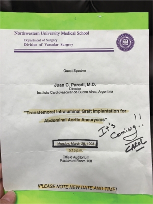
Figure 1: Announcement of Dr. Parodi's keynote lecture at the Northwestern Vascular Symposium in 1993
An oral history interview with Jon S. Matsumura, MD adds a substantial claim, noting that Northwestern faculty served as principal investigators on all three of the earliest FDA-approved aortic endograft trials in the country. By Matsumura's account, Yao led the EVT (later Ancure) trial. Pearce, who had served as a paid Medtronic advisor and recused himself to avoid conflict of interest, asked Matsumura to serve as PI for the Medtronic AneuRx trial in his place. Matsumura served separately as national PI for the Gore Excluder trial. This places Northwestern's faculty simultaneously embedded in the leadership of the entire first generation of FDA-approved aortic endografts, rather than as an early adopter of a single device (Figure 2).
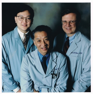
Figure 2: Pictured from L to R: Dr. Jon Matsumura, Dr. James Yao, Dr. William Pearce, Northwestern Vascular Surgery faculty and national PIs of the early aortic stent graft clinical trials
The First Endograft and the Device Learning Curve (1993 - Present)
Pearce dated Northwestern's first aortic endograft precisely December 1993. An EVT device was placed by Pearce and Yao for an abdominal aortic aneurysm. Pearce recalled that this was the first endograft placed in Chicago. Pearce was candid that the earliest devices were close to experimental. The EVT device required an open femoral or iliac exposure to accommodate an oversized introducer sheath, fixation hooks were found to fracture over time, and many planned cases were converted to open repair when the device could not be advanced. The second-generation Medtronic AneuRx graft brought its own problems. Early grafts, hand-sewn to their metal exoskeleton, developed microleaks at nearly every suture line, causing persistent sac pressurization now recognized as endotension. Before placing early grafts of this kind, Pearce and Yao traveled to a large-animal laboratory in Utah to rehearse implantation on cattle-sized animals. Dr. Eskandari's own interview recounts a similar rehearsal of Yao, Pearce, and Vogelzang practicing stent-graft deployment on cows sometime in the mid-to-late 1990s, before Eskandari himself joined as a fellow. Whether this was the same Utah trip or a separate occasion remained unresolved in both interviews. Regardless, both accounts validate that live-animal practice was a deliberate, repeated practice of how the division prepared to integrated a new technology, rather than a one-time event.
Building a Graft from Scratch: The First Hand-Made TEVAR Endograft
A case presentation prepared by Melina R. Kibbe, MD documents a still more improvisational chapter in this history, involving a thoracic endograft hand sewn on the back table when no commercial device existed to fit a patient's anatomy. In April 2003, a team including Drs. Matsumura, Eskandari, and Kibbe treated a descending thoracic aortic aneurysm using a custom endograft. Two hand-sewn Z-stents mounted on an ironed Dacron graft were assembled at the back table and loaded into an improvised delivery system with successful exclusion of the pseudoaneurysm confirmed on completion angiogram (Figure 3).
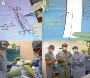
Figure 3 A-D: Construction of hand made TEVAR stent graft by connecting two 40mm x 5cm Z stents (A) to a 38mm ironed dacron graft (B). Back table reconstruction by Drs. Mark Eskandari and Melina Kibbe (C), and surgical team after graft implantation Drs. Jon Matsumura, Mark Eskandari, and Melina Kibbe (D)
Eskandari confirmed in his own oral history interview that this was not an isolated case but one of a working series. He recounted that the team built and implanted around fifteen to twenty such custom thoracic devices before commercially manufactured thoracic endografts became available. Eskandari credited Matsumura with originating this culture of innovation at Northwestern. Matsumura separately recalled building comparable "homemade" abdominal endografts as early as 1999–2000, suggesting the 2003 thoracic case continued an already-established practice. The series was formally published as Peterson, Longo, Matsumura, Kibbe, Morasch, Cardeira, and Eskandari, "Endovascular Repair of Thoracic Aortic Pathology with Custom-Made Devices," in Surgery in 2005.
CT, Objective Measurement, and the Hybrid Operating Room
A further less celebrated Northwestern contribution was the early adoption of computed tomography for aortic disease at a time when CT's value for this purpose was not widely accepted, even within the specialty. Vogelzang described demonstrating CT's utility directly to Drs. Bergan, Yao, and Flynn. The clinical case for the switch was strongest for aortic dissection, which he described as effectively impossible to characterize reliably on catheter angiography. The transition also carried a safety benefit. Before CT, aortic anatomy was commonly evaluated with translumbar aortography; a direct lumbar puncture of the aorta performed hundreds of times over the years. Only once CT became routine did the group fully appreciate how often that puncture had produced clinically significant retroperitoneal hemorrhage.
Matsumura traced a recurring theme across the division's diagnostic and research history - completion angiography, routine postoperative ankle-brachial index testing, and a dedicated aortic imaging core laboratory - to James Yao's insistence on objective, quantifiable measurement over clinical impression alone. As a fellow in the mid-1990s, Matsumura was sent to independently review imaging for the first FDA trial of an aortic endograft. He co-authored what became the first published demonstration that an aneurysm could shrink after endovascular repair without open surgery (Matsumura, Pearce, McCarthy, and Yao, Journal of Vascular Surgery, 1997). That body of work also documented the phenomenon now called "endoleak," seeding further Northwestern outcomes research, including the division's contributions to percutaneous access technique (Morasch, Kibbe, Evans, Meadows, Eskandari, Matsumura, and Pearce, Journal of Vascular Surgery, 2004).
The eventual creation of a combined operative and angiographic suite, the hybrid OR, now considered a baseline requirement for any vascular surgery and complex aortic program, followed a similar arc from improvisation to formal infrastructure. According to Pearce, the division initially relied on a hanging portable x-ray unit simply to obtain completion angiograms after open surgery. The very first endograft was placed using this improvised setup. After the move to the new Northwestern Memorial Hospital building in 1999, Pearce and Yao secured philanthropic funding and institutional support to build a dedicated angiography suite directly into an operating room, Room 26, over some resistance from interventional radiology, which Pearce described in frankly territorial terms, "They didn't want us doing this stuff." Matsumura's interview independently substantiated and extended this account, specifying the Philips V5000 fluoroscopy unit installed and recalling early design flaws (equipment mounted from the ceiling that staff repeatedly struck their heads on). Later hybrid-OR designs elsewhere corrected these flaws. Pearce and Matsamura both corroborated that Northwestern was among the first, if not the first center, to install a Phillips fixed imaging system in an operating room, and served as a blueprint for the development of hybrid operating rooms around the world.
The Hand-Sewn Tradition Continues
The back table improvisation documented earlier in this chapter continued beyond the early 2000s. Photographs taken by the author in 2014 show Andrew W. Hoel, MD, and Tadaki M. Tomita, MD, hand-constructing a physician-modified endograft (PMEG), a commercially manufactured stent graft laid out on the back table with fenestrations or branch sites tailored to a specific patient's renal and visceral artery anatomy (Figure 4). Eskandari dated the division's first fenestrated case to 2012 or 2013, with Dr. Hoel was recruited specifically to build that practice.
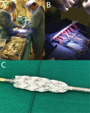
Figure 4: Back table modification of a thoracic aortic endograft for use in the viseceral segment aorta by Drs. Andrew Hoel and Tadaki Tomita (A); Thoracic aortic endograft reconstrained for replacement in its original delivery system after back table customization (B); Infrarenal bifurcated aortic endograft modified for use in the visceral segment aorta (C)
Physician-modified endografting emerged nationally in the early-to-mid 2000s to treat complex aneurysms in patients whose anatomy was not suitable for an off-the-shelf device. The current practice extends this thread further, with continued physician-modified fenestration and branch construction alongside newer, purpose-built platforms (Gore TAMBE and Cook Zenith ZFEN+ systems). The tradition of objective completion assessment further continues today with routine use of cone-beam CT rather than two-dimensional completion angiography alone (Figure 5).
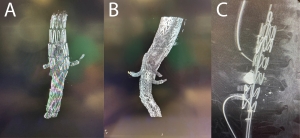
Figure 5: Completion cone beam CT used as objective measurement of satisfactory complex endovascular aortic reconstruction using a Cook Zenith Fenestrated Plus device (A); Gore Thoracoabdominal Branch Endoprosthesis (B); and physician modified Cook Zenith Alpha (C)
That endovascular ascendance has a necessary counterweight in cases where a prior endograft fails years later with endoleak or infection, requiring full explantation and conversion to open thoracoabdominal repair. Eskandari described this, not as an occasional correction, but as a purposeful institutional stance. When asked about the biggest internal disagreements as the division moved from open to endovascular repair, he pointed to a collective wariness about pushing endovascular therapy past its proven limits, describing Northwestern's faculty as "pretty conservative" as a group, having watched "many, many grafts that came and went that sounded great, but didn't last very long." That open thoracoabdominal expertise, inherited from Bergan, Yao, and Pearce's generation, remains an active clinical practice, rather than a historical footnote to the endovascular era (Figure 6).
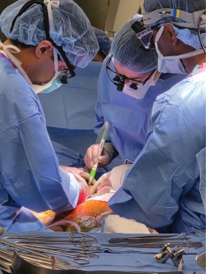
Figure 6: Open extent IV thoracoabdominal aortic reconstruction for failed aortic endograft performed by Northwestern integrated vascular surgery alum Dr. Neel Mansukhani, and current integrated vascular surgery residents Drs. Margaret Reilly and Shaunak Roy
The Present: A Multidisciplinary Aortic Program
Today the Division of Vascular Surgery is led by Mark K. Eskandari, MD, FACS, the James S. T. Yao, MD, PhD, Professor of Vascular Surgery and System Chief of the Division, whose own research has centered on thoracic and abdominal endovascular aortic repair, including several significant early published series that helped establish TEVAR as first-line therapy for traumatic and ruptured thoracic aortic disease, and later contributed directly to the SVS treatment guidelines for Management of Abdominal Aortic Aneurysm, and Management of Blunt Traumatic Aortic Injury. Dr. Eskandari trained under Dr. Yao and later carried that tradition of education and mentorship forward through his leadership of the division. In 2009, he and Dr. Heron Rodriguez established the Northwestern Vascular Surgery Integrated Residency Program, one of the first integrated vascular surgery training programs in the country.
The division's aortic work is now organized within the Vascular Surgery Comprehensive Aortic Program (CAP) and takes a patient-centered approach to degenerative aneurysms, dissection, connective tissue and heritable aortopathies, aortic infection, and complex reoperative anatomy. Until recently, Andrew W. Hoel, MD, directed the CAP and served as the division's Director of Outcomes and Quality Improvement. This established cross-departmental structure can be viewed as the institutional descendant of the informal collaboration between vascular surgery and interventional radiology described above. What began as personal relationships between a handful of physicians has become deliberately incorporated within the structure of the program itself.
A defining feature of the current program is its integration with the Bluhm Cardiovascular Institute’s cardiac surgery team. Within the Bluhm Cardiovascular Institute, the Comprehensive Aortic Center, which integrates vascular and cardiac surgery, cardiology, cardiovascular genetics, and radiology extend the aortic program’s reach to the aortic root, arch, and connective tissue disease, areas at the boundary between cardiac and vascular surgery. Dr. S. Chris Malaisrie performed one of the first procedures in Illinois using an investigational "frozen elephant trunk" hybrid graft for combined arch and descending aortic repair, a technique that has since become a common practice at Northwestern. Together, the divisions of vascular surgery and cardiac surgery have implemented a direct-to-operating-room transfer pathway for acute aortic emergencies under the leadership of Dr. Christopher K. Mehta.
Today, the vascular surgery division performs aortic surgery for aneurysms, dissection, occlusive disease, and trauma on a daily basis. If a patient is treated with open surgery, endovascular therapy, or hybrid techniques depends entirely on what is best for the patient. Strategies are implemented to minimize radiation exposure to patients and providers, novel technologies are integrated monthly, and techniques are under constant refinement. System wide and multidisciplinary aortic conferences are held twice monthly, and formal and informal multidisciplinary collaborative relationships are stronger than ever before. Northwestern faculty continue to participate in every current major aortic clinical trial, and present contributions in the form of innovation and investigation related to aortic diseases at every major conference. The expertise of aortic surgeons at Northwestern earns invitations at meetings locally, regionally, nationally, and internationally every year.
Imaging Innovation: 4D Flow MRI
Northwestern's aortic program has also been shaped by a research strength unusual for a vascular program in advanced cardiovascular imaging physics. Michael Markl, PhD, and Bradley Allen, MD are internationally recognized as pioneers of four-dimensional (4D) flow MRI, a technique that captures time-resolved, three-directional blood flow throughout the aorta. 4D flow MRI is currently used to characterize abnormal wall shear stress in bicuspid aortic valve-associated aortopathy, to track hemodynamic changes in type B aortic dissection over time, guide therapies for type B aortic dissection, and to develop automated processing pipelines intended to bring these measurements into routine clinical use. This work speaks directly to an unmet need raised independently: the need for a better biological understanding of which aneurysms and dissections actually threaten a given patient (Figure 7).
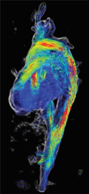
Figure 7: 4D MRI of a subacute uncomplicated type B dissection in a lateral projection
Training the Next Generation
The division's educational mission remains central today. Northwestern's vascular surgery training now includes both a traditional fellowship (5+2) and an integrated residency pathway (0+5), coordinated with the McGaw Medical Center of Northwestern University. Established by Bergan and Yao in 1974–1975 as one of the earliest formal vascular fellowships in the country, the program has grown from training a single fellow at a time to roughly twelve trainees a year. Yao alone is credited with having personally trained more than sixty vascular fellows. Currently, the division of vascular surgery is pioneering a ruptured aortic aneurysm life support training program, modeled after advanced cardiovascular life support (ACLS) to systematically train providers in protocolized and structured management of patients presenting with this acute life threatening disease.
The Future: Where the Program Is Headed
Several developments suggest how Northwestern's aortic surgery practice may evolve over the coming decade. Further collaboration between vascular and cardiac aortic teams, through the Comprehensive Aortic Program and Bluhm's Comprehensive Aortic Center, is likely to deepen for arch and thoracoabdominal pathology that does not respect the traditional boundary between the two specialties. Complex endovascular techniques such as fenestrated and branched endografting are likely to continue displacing open repair for anatomically complex aneurysms in appropriately selected patients, even as open thoracoabdominal and arch surgery remain essential for younger patients, connective tissue disease, and anatomy unsuited to current devices.
Building on Northwestern's 4D flow MRI program and its growing use of artificial intelligence to automate hemodynamic analysis, imaging and data-driven risk stratification may allow earlier, more individualized identification of patients whose aortic disease is likely to progress, rather than relying primarily on diameter-based thresholds. This direction was echoed strikingly in oral history for this chapter: In discussing the field's greatest unmet need, Robert Vogelzang pointed not to a new device but to basic biology, specifically the incompletely understood natural history of aortic aneurysms and the concern that current practice may risk "treating pictures, not patients." He argued that modern imaging finds abnormalities so readily that clinicians can be tempted to intervene on an incidental finding rather than a genuine risk of rupture. He predicted that the current volume of infrarenal aneurysm repair may, in retrospect, be seen as having been driven partly by the availability of effective technology rather than a fully mature understanding of which aneurysms truly threaten a given patient.
The current Comprehensive Aortic Program’s vision that guides current efforts is that no one should suffer from aortic disease. As such, we envision a future where a patient’s anxiety surrounding aortic diseases is manageable to non-existent; radiation exposure in the course of aortic disease diagnosis, surveillance, and treatment, is less than that of a cross country flight; aortic surgery is done as on an outpatient basis; and spinal cord ischemia and paralysis from aortic surgery will be nothing more than a bad memory of seemingly barbaric treatment strategies of the past. The division's continued investment in outcomes measurement, quality improvement, and its own trainee program suggests that Northwestern's aortic program intends to keep producing not only excellent individual outcomes but the next generation of surgeon-scientists who will carry this work forward, as Northwestern alum have done for the past fifty years.
Patient Care
Comprehensive clinical care of aortic disease is a core element of our Program. For more information about our clinical expertise and team, please see our Northwestern Medicine page via the link below.
Research
The CAP has a mandate to advance the optimal care and treatment of aortic disorders. This encompasses both understanding the pathophysiology and natural history of aortic disorders, advancing the “technology and techniques” for treatment and understanding the impact of aortic disease and disease treatment for patients and their family/caregivers. We will:
- Participate in multiple clinical trials advancing the minimally invasive treatment of complex aortic disease. This will be in collaboration with industry partners and the BCVI Clinical Trials Unit.
- Advance institutionally sponsored clinical trials advancing the minimally invasive treatment of complex aortic disease.
- Leverage developing translational research infrastructure (tissue biobank) to investigate pathophysiology of aortic disease and potential role for microbiome.
- Utilize EDW and retrospective clinical data review to improve our clinical care processes, optimize collaboration with other specialties and to disseminate our experience.
- Continue to engage with VQI and VISION to leverage large data to expand our understanding of aortic disease, device innovation and appropriate use.
- Expand our qualitative research structures to support of our efforts around patient education and appropriate use.
Current studies being undertaken by the CAP
Study Name: LumiGuide Xperience Registry
- Prospective, observational (real-world), multi-center study of intraoperative aortic patients undergoing endovascular aortic repair.
- Philips Lumiguide device system, powered by Fiber Optic RealShape (FORS) technology, uses light to provide real-time, 3-D, full-shape visualization of guidewires and catheters as surgeons navigate arterial vasculature during complex endovascular aortic procedures.
- Yields a reduction in intraoperative fluoroscopy time and radiation dose.
1.
Video 1: Barrel View Cannulation
2.
Video 2: Celiac Cannulation
Study Name: IMPRoving Outcomes in Vascular DisEase – Aortic Dissection (IMPROVE-AD) Trial
- Prospective, pragmatic, randomized, multi-center clinical trial of patients with a subacute uncomplicated type B aortic dissection.
- Objective: Determine whether upfront TEVAR plus medical therapy reduces all-cause mortality or major aortic complications compared with medical therapy and surveillance for patients with subacute uncomplicated type B aortic dissection.
Study Name: Serial Examination of Abdominal Aortic Aneurysm Patients with Computed Tomography
- Prospective, observational, multi-center study of patients with a small AAA managed with medical surveillance for three years.
- Apply a physics-based method for predicting future AAA growth by calculating RPI through use of annual abdominal contrast-enhanced or unenhanced CT exams.
Study Name: Gore TBE Registry
- Post Market, prospective, non-randomized, multi-center, observational study.
- Patient Population: Aortic patients who undergo treatment with the GORE® TAG® Thoracic Branch Endoprosthesis device.
- Objective: To ensure that the clinical outcomes during the commercial use of the GORE® TAG® Thoracic Branch Endoprosthesis are as anticipated.
Study Name: Treatment of Post-EVAR Endoleak using SHApe Memory Polymer: A Multicenter Evaluation (TOP SHAPE)
- Multicenter international retrospective registry.
- Patient population: Patients undergoing reintervention or embolization for post-endovascular type II endoleak using Shape Memory Polymer (SMP) embolization plugs.
Zenith fenestrated plus: Evaluation of a novel stent graft for the endovascular treatment of thoracoabdominal and juxtarenal abdominal aortic aneurysms
ARISE II: Evaluation of the GORE® Ascending Stent Graft in the Treatment of Chronic Lesions of the Ascending Aorta
ARISE III: Evaluation of the GORE® Ascending Stent Graft in the Treatment of Acute Lesions of the Ascending Aorta
Education
We apply a holistic approach to the educational endeavors of the CAP acknowledging that patients, physicians, physicians-in-train, nurses, clinical staff, and administration can all contribute to the teaching and learning environment of the program.
- Patient-centered care and education is a hallmark of the CAP. This includes disease and procedure education, education related to heritability of aortic disorders, risk stratification education, and decision support.
- Expansion of education opportunities will target Northwestern trainees in the Division of Vascular Surgery and the Department of Surgery. In collaboration with the broader NM and NU community, educational opportunities will additionally focus on trainees, staff, and faculty in NM and NU departments involved in the treatment of patients with aortic disease.
- Outreach will engage regional clinical providers to advance care quality care delivery to patients with aortic disease. This will focus on perioperative and longitudinal care of patients with aortic disorders.
- Surgeon education within and beyond NM will focus on diagnosis and treatment of aortic disorders, particularly related to advanced endovascular therapy and clinical decision-making.
Vascular Aortic Conference
- Monthly system-wide Division of Vascular Surgery conference.
- Team discussion of complex patient cases for treatment and surgical case planning, as well as programmatic updates.
Multidisciplinary Aortic Conference
- Monthly conference with multidisciplinary team, including vascular surgery, cardiac surgery, cardiology, cardiovascular genetics, and radiology.
- Discussion of shared complex patient cases for treatment and surgical planning, as well as programmatic updates.
Code Aorta Type B Aortic Dissection Bedside Education
- Aortic RN coordinator provides bedside aortic dissection and blood pressure education to inpatients with new type B aortic dissection prior to discharge. Home BP device and aortic dissection/blood pressure education packet provided to patients for long-term home use.
- Multidisciplinary outpatient follow-ups scheduled prior to discharge by inpatient vascular APP team and aortic RN coordinator.
- Aortic RN coordinator surveils patients with new type B dissection as outpatients.
Patient Education Brochures
- AAA and TAA/TAAA patient education brochures developed by team to provide to patients and their support persons.
Contact Us

Brianna Martinek, BSN, RN
Aortic RN Coordinator